Rheumatoid Forefoot Deformity
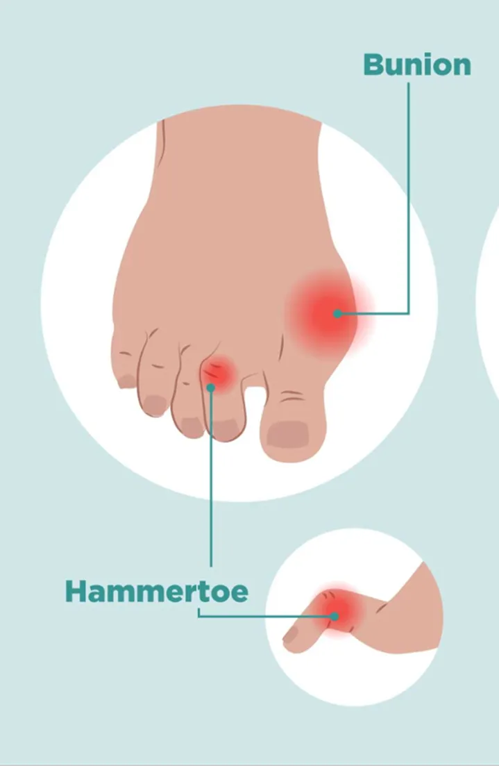
Rheumatoid forefoot deformity develops when long-standing inflammation affects the joints at the ball of the foot. Ongoing swelling inside the metatarsophalangeal (MTP) joints gradually damages cartilage, bone, and the supporting ligaments and tendons. As this support weakens, the smaller toes drift upward and out of alignment, and the natural cushioning under the ball of the foot shifts away from its normal position. This leaves the heads of the bones more exposed to pressure, often causing painful calluses and discomfort when walking. At the same time, the big toe frequently drifts outward (hallux valgus), which reduces its ability to bear weight and transfers more load to the central forefoot. Together, these changes can widen the forefoot, reduce push-off strength, and make standing and walking increasingly painful even when the active inflammation is controlled with medications.1,2
The Procedure
Rheumatoid forefoot reconstruction is designed to relieve pain, correct deformity, and improve the way the foot bears weight during standing and walking. The procedure is tailored to the pattern and severity of deformity but most commonly involves correcting the big toe joint and addressing the painful joints under the ball of the foot:
● The big toe joint is often stiff, damaged, or unstable. In this situation, the joint is surgically fused in a straight, functional position to create a stable first ray that can reliably bear weight and help redistribute pressure away from the central forefoot.
● The lesser toes can also be painful; to address this, the painful ends of the metatarsal bones are often shortened or removed to reduce pressure under the foot and allow the toes to realign.
● Toe deformities (e.g. claw toe, hammertoe) can be associated; they are corrected at the same time using soft-tissue releases, small bone procedures, or temporary pins to hold the toes straight during healing.
In selected patients with less severe joint damage, joint-preserving techniques may be used instead of removing bone. These approaches aim to realign the toes while maintaining more of the natural joint structure, but they are used selectively and depend on bone quality, deformity severity, and overall disease control.1,2
Risks and Complications
Rheumatoid forefoot reconstruction is generally associated with significant improvements in function, pain, and quality of life. However, as with any surgery, complications can occur. The most common ones consist of:
● Delayed wound healing (≈ 20%): slower-than-expected healing of the surgical incision
● Recurrence of hallux valgus (≈ 7.5–32%): return of outward deviation of the big toe
● Hallux varus deformity (≈ 15%): overcorrection causing inward deviation of the big toe
● Recurrence of lesser toe MTP subluxation/dislocation (≈ 11%): return of instability or dislocation of the smaller toe joints
● Nonunion (≈ 0–26%): incomplete bone healing, often without symptoms
● Reoperation (< 15%): need for additional surgery due to complications or persistent deformity 2-6
The rates of each complication vary depending on the surgical technique and patient factors. As for other surgeries, general risks related to surgery and anesthesia may include infection, bleeding, blood clots, allergic reactions, cardiopulmonary complications, and wound-healing issues. Any other general surgical and anesthesia-related risks will be discussed with you prior to surgery if applicable.
What to Expect
Before surgery, your surgeon will examine your foot and review imaging (e.g. foot x-rays) or diagnostic tests if needed to assess the extent of the disease and joint involvement. Rheumatoid forefoot reconstruction is usually performed under regional anesthesia (such as a nerve block), sometimes with sedation, or under general anesthesia, often depending on patient-specific factors.7 After surgery, your foot will be dressed and protected in a postoperative shoe, and pain is usually managed with acetaminophen and anti-inflammatory medications.
Your Recovery
Patients usually return home the same day following rheumatoid forefoot reconstruction, and progressive, protected weightbearing is typically allowed as tolerated after the surgery. Keeping the foot elevated during the first one to two weeks helps reduce swelling and promote healing, and stitches are typically removed after about two weeks.
During the first 6 weeks, the main goals are wound healing and maintaining proper alignment. X-rays are typically obtained by 2 months to confirm bone healing and stability. Provided the foot is healing well, many patients begin transitioning to regular footwear and activities between 2 and 3 months, while the return to full unrestricted activities typically occurs by 4 months. Functional improvement typically continues up until the first year following surgery. 2-4,8-9
Throughout your healing journey, follow-up visits with your surgeon ensure proper healing and guide your safe return to activity at each stage of recovery.
References
Louwerens JW, Schrier JC. Rheumatoid forefoot deformity: pathophysiology, evaluation and operative treatment options. Int Orthop. 2013;37(9):1719-1729. doi:10.1007/s00264-013-2014-2
Whitt KJ, Rincker SA, Hyer CF. Sustainability of Forefoot Reconstruction for the Rheumatoid Foot. J Foot Ankle Surg. 2016;55(3):583-585. doi:10.1053/j.jfas.2016.02.003
Yano K, Ikari K, Tobimatsu H, Okazaki K. Patient-Reported and Radiographic Outcomes of Joint-Preserving Surgery for Rheumatoid Forefoot Deformities: A Retrospective Case Series with Mean Follow-up of 6 Years. J Bone Joint Surg Am. 2021;103(6):506-516. doi:10.2106/JBJS.20.01144
Etani Y, Hirao M, Ebina K, et al. Combination of Modified Scarf Osteotomy and Metatarsal Shortening Offset Osteotomy for Rheumatoid Forefoot Deformity. Int J Environ Res Public Health. 2021;18(19):10473. Published 2021 Oct 5. doi:10.3390/ijerph181910473
Takakubo Y, Wanezaki Y, Oki H, et al. Forefoot Deformities in Patients with Rheumatoid Arthritis: Mid- to Long-Term Result of Joint-Preserving Surgery in Comparison with Resection Arthroplasty. Int J Environ Res Public Health. 2021;18(21):11257. Published 2021 Oct 26. doi:10.3390/ijerph182111257
He Y, Shan F, Fan C, Zeng X, Yang G, Tang B. Effectiveness of the First Metatarsophalangeal Joint Arthrodesis Versus Arthroplasty for Rheumatoid Forefoot Deformity: A Systematic Review and Meta-Analysis of Comparative Studies. J Foot Ankle Surg. 2021;60(4):787-794. doi:10.1053/j.jfas.2020.06.031
Lee TH, Wapner KL, Hecht PJ, Hunt PJ. Regional anesthesia in foot and ankle surgery. Orthopedics. 1996;19(7):577-580.
Shimomura K, Yasui T, Teramoto A, Ozasa Y, Yamashita T, Iwasawa M. Time Course of Quality of Life Improvement Between Resection Arthroplasty and Metatarsophalangeal Joint-Preserving Forefoot Arthroplasty for Rheumatoid Arthritis. Foot Ankle Int. 2021;42(2):166-175. doi:10.1177/1071100720962096
Triolo P, Rosso F, Rossi R, Cerlon R, Cottino U, Bonasia DE. Fusion of the First Metatarsophalangeal Joint and Second to Fifth Metatarsal Head Resection for Rheumatoid Forefoot Deformity. J Foot Ankle Surg. 2017;56(2):263-270. doi:10.1053/j.jfas.2016.11.008
.svg)

.svg)
.svg)
.svg)
.svg)
